
Bookmark this lesson
Login or create an account to save and access your bookmarks
Bookmark this lesson
Login or create an account to save and access your bookmarks
Not yet registered?
What you can access by simply creating a account:
Register today to get the most out of the education academy. Save your progress, bookmarks lessons, track your progress plus more...

Save your progress in this lesson so you can pick it up where you left off.

Add lessons to your bookmarks so you can quickly return to them in the future.

Track your progress throughout the course to see what you have achieved.
Organ affected: lungs
IgG4-RD can affect the lungs quietly. Learn about symptoms, diagnosis, treatments and long term management.
 6 lessons
 Total: tbc
Organ overview
How IgG4-RD can affect the lungs
When you hear that IgG4-related disease, or IgG4-RD, may involve the lungs, it can feel frightening. The lungs are tied to something we do every moment: breathing. So even before we talk about scans or tests, it helps to begin with the basics. What do the lungs do? What are their main parts? And where might IgG4-RD show up?
Keep in mind that lung involvement does not look the same for everyone. Some people have cough, wheezing, shortness of breath, or chest pain. Other people have no symptoms, and lung changes are noticed only because imaging was done for another reason. When lung disease is identified by chance or accident, so to speak, the X-ray or CT results are often termed “incidental findings” by medical professionals. Those “incidental findings,” however, can have big implications (and often cause great worry) for patients.
Lesson 1 gives you an overview of IgG4-RD affecting the lungs. Later lessons will explain evaluation, treatment, and monitoring in more detail.
IgG4-RD and lung involvement
In this short video clip, Dr. Jason Springer explains the basic parts of the lungs and the different ways IgG4-RD can affect lung tissue, airways, lymph nodes, and the lining around the lungs.
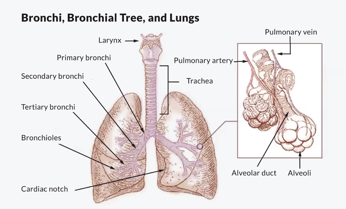
Parts of the lung and related areas
Trachea | Main tube that brings air into the chest. |
|---|---|
Bronchi | Branches that carry air into each lung. |
Bronchioles | Smaller branches move air deeper into the lungs. |
Alveoli | Tiny filters that clean the blood. |
Pleura | Thin lining that can become thickened or irritated. |
Mediastinum | Middle chest area where lymph nodes may enlarge. |
Hilum | Place where airways and blood vessels enter. |
Lymph nodes | Immune filters may enlarge when disease is active. |
The simple map of the lungs
The lungs are soft, sponge-like organs with a delicate framework that supports the air sacs and blood vessels. This framework is sometimes called the interstitium. The lungs bring oxygen into the body and remove carbon dioxide. Air enters through the trachea, or windpipe, then moves into two large breathing tubes called bronchi. These branch into smaller tubes called bronchioles. At the ends are tiny air sacs called alveoli. It is through the walls of the alveoli that oxygen moves into the blood, where it can be pumped by the heart to the rest of the body.
Between the lungs is the mediastinum, a central chest space that contains lymph nodes and the body’s biggest blood vessels (for example, the aorta). The lungs also have a thin outer lining called the pleura. IgG4-RD can sometimes affect both of these regions, as well. If the mediastinum is involved, the condition is often called “fibrosing mediastinitis”. When the pleura are involved by inflammation, the term “pleuritis” is used.
What the lungs do
The lungs have one main job: they help move oxygen into the body and carbon dioxide out of the body. When you breathe in, oxygen travels deep into the lungs. From there, oxygen passes into the blood so it can be carried to the brain, heart, muscles, kidneys, and every other part of the body. When you breathe out, carbon dioxide leaves the blood and exits through the lungs. The lungs and the system in which they reside are a remarkable physiological machine!
If IgG4-RD affects the airways, a person may notice cough or wheeze. If it affects the air sacs or the tissue around them, a person may notice shortness of breath or fatigue. Sometimes, though, the changes are mild enough that a person does not notice symptoms at all. Most patients with IgG4-RD involving the lung have almost no symptoms for a long time, even in the setting of what is sometimes rather extensive lung disease.
— Dr. Leigh Minuk, respirologist/pulmonologist, Vancouver General Hospital / Centre for Lung Health, Vancouver, BC
In this video, Dr. Leigh Minuk, a pulmonologist/respirologist from Vancouver General Hospital, explains that lung involvement in IgG4-RD may be under-recognized.
Where IgG4-RD can appear in the chest
IgG4-RD can affect different parts of the lungs and chest. It may involve the lung tissue, the airways, the blood vessels, the pleura, lymph nodes, or the mediastinum.1
In this video, Dr. Jason Springer, rheumatologist, Associate Professor of Medicine, Vanderbilt University Medical Center, explains how IgG4-RD can affect the lungs.
What doctors may see:
A nodule or mass, meaning a small or larger spot of inflammation within the lung.
A ground-glass opacity, meaning a hazy area on the scan. An area of “ground-glass” is typically less solid than a nodule or mass.
An interstitial change, meaning the lung’s support tissue is involved.
An airway change, meaning breathing tubes look thickened or narrowed.
Pleural thickening, meaning the lining around the lung looks thicker.
Enlarged lymph nodes, meaning immune-system filters in the chest are bigger than expected. The lymph nodes are sites within the body where cells of the immune system mature normally. In times of inflammation, it is natural for lymph nodes to become bigger (there are more cells in them).
Sometimes IgG4-RD can cause fibrosis, which means scar-like tissue. In the lungs, fibrosis may involve the support tissue around the air sacs.
— Dr. Jason Springer, rheumatologist, Associate Professor of Medicine, Vanderbilt University Medical Center
In this video, Dr. Jason Springer, co-director of the Vanderbilt Vasculitis Center at Vanderbilt University Medical Center, talks about how IgG4-RD can appear in the lungs.
Lung IgG4-RD can mimic other conditions
One challenge is that lung IgG4-RD can look like other lung diseases. A nodule or mass can raise concern for cancer. A hazy area can look like infection or inflammation. Airway changes can feel like asthma. Interstitial changes can resemble other forms of lung scarring.
This does not mean the finding is cancer, infection, or asthma. It means doctors have to be careful. IgG4-RD is one possible explanation, but it is not the only one.
That is why diagnosis usually depends on the full picture: symptoms, imaging, blood tests, other organ involvement, and sometimes tissue from a biopsy.
The main tests doctors may use
A chest X-ray may be used as a first look, especially when a person has cough, chest pain, or shortness of breath. But X-rays may miss smaller or more subtle lung findings.
A CT scan gives a much more detailed view of the lungs. It can show nodules, hazy areas, airway thickening, pleural thickening, enlarged lymph nodes, and interstitial changes. For lung IgG4-RD, CT is often one of the most useful imaging tools. One important caveat, though: CT scans are very sensitive and often turn up “shadows” that are really nothing to worry about. So not every shadow that shows up on a scan or in a radiology report represents pathology (i.e., something of potential concern).
A PET scan may show whether an area is active. Active areas can include inflammation, infection, or cancer, so PET scans can be helpful but not definitive. They show where activity is happening (and, like CT scans, are very sensitive, sometimes overly so). But they seldom offer definitive clues about WHY the inflammation is happening, or even, in fact, whether the inflammation is associated with IgG4-RD, with infection, or with cancer. The precise WHAT and WHY have to be interpreted by the clinician in context—a process that is often termed “clinico-radiologic correlation.”
Pulmonary function tests, or PFTs, are breathing tests. They measure how well air moves in and out of the lungs and how well oxygen moves from the lungs into the blood. PFTs can help show whether lung findings are affecting breathing function.
When biopsy becomes important
A biopsy is a small tissue sample that doctors study under a microscope. In lung IgG4-RD, biopsy may be needed when imaging cannot clearly explain what is happening, especially if a nodule or mass looks concerning. Biopsies are often needed to clinch an IgG4-RD diagnosis, and are also necessary sometimes to exclude cancer, infection, or other processes.
There are different ways to sample lung tissue. Some biopsies use a needle. Some are done through a camera passed into the airways. Some require a surgical approach to collect a larger tissue sample. The choice depends on where the abnormal area is, how much tissue is needed, and what is safest for the patient.2
Pathologists then look for features that support IgG4-RD, including IgG4-positive plasma cells, inflammation, and fibrosis. But even biopsy results must be interpreted in context. A tissue sample is one important clue, not a stand-alone answer.
Why other organs matter
IgG4-RD often affects more than one organ. The correct interpretation of abnormalities in the lung findings may make more sense if a person is known to have IgG4-RD involvement in the pancreas, bile ducts, salivary glands, eyes, kidneys, aorta, or lymph nodes.
This is why doctors may ask about symptoms outside the lungs. They may also review prior scans, blood tests, and biopsy results from other areas of the body. Sometimes the lung is the first clue–the organ that points ultimately to the correct diagnosis. Other times, lung findings are part of a broader IgG4-RD pattern.
How the diagnosis comes together
Diagnosing lung IgG4-RD is often a team process. A pulmonologist focuses on breathing, lung imaging, and lung tests. A rheumatologist looks at the whole-body immune disease pattern. A radiologist studies the imaging. A pathologist studies tissue if a biopsy is done.
The team may ask:
Does the CT pattern fit IgG4-RD?
Could this be infection, cancer, asthma, sarcoidosis, vasculitis, or another lung disease?
Are other organs involved?
Do blood tests support the picture?
Is biopsy needed to clarify the diagnosis?
Does the biopsy show findings that fit IgG4-RD?
The goal is to avoid both underdiagnosis and overdiagnosis. IgG4-RD can be missed, but it can also be mistaken for other conditions. Careful diagnosis helps guide the right next steps.
Summary
IgG4-RD can involve several areas of the lungs and surrounding tissue. It may appear as nodules, masses, ground-glass opacities, interstitial changes, airway changes, pleural thickening, enlarged lymph nodes, or mediastinal involvement. Diagnosis is not based on tests or imaging alone. Doctors combine symptoms, CT findings, PET findings when needed, pulmonary function tests, blood tests, other organ involvement, and sometimes biopsy. The goal is to understand whether the lung findings are truly part of IgG4-RD or better explained by another condition.
References
Springer J. IgG4-RD lung breakout session. IgG4ward JAM. https://igg4ward.org/resources/igg4ward-jam-video-series-video-3-igg4-rd-and-the-lungs/
Minuk L. The lungs and IgG4-RD. IgG4ward CanJAM.
Muller R, Ebbo M, Habert P, Daniel L, Briantais A, Chanez P, Gaubert JY, Schleinitz N. Thoracic manifestations of IgG4-related disease. Respirology. 2023;28(2):120–131. doi:10.1111/resp.14422. https://pmc.ncbi.nlm.nih.gov/articles/PMC10100266/
Stone JH, Zen Y, Deshpande V. IgG4-related disease. New England Journal of Medicine. 2012;366(6):539–551. doi:10.1056/NEJMra1104650. https://www.nejm.org/doi/full/10.1056/NEJMra1104650
 Save this lesson so you can resume it later
 Copy Link
 Share On X
 Share On Facebook
 Share On LinkedIn
 Share in Whatsapp
Further reading
IgG4ward CanJAM 2025 National Heart, Lung, and Blood Institute overview of how the lungs work Explanation of chest CT imaging from RadiologyInfo.orgPatient guides for doctor appointments
Educating new doctors and healthcare professionals on your IgG4-RD journey can feel like a full-time job. Download, print, or have your doctor scan the QR code of our patient guides to make every appointment more productive.
View guides