
Bookmark this lesson
Login or create an account to save and access your bookmarks
Bookmark this lesson
Login or create an account to save and access your bookmarks
Not yet registered?
What you can access by simply creating a account:
Register today to get the most out of the education academy. Save your progress, bookmarks lessons, track your progress plus more...

Save your progress in this lesson so you can pick it up where you left off.

Add lessons to your bookmarks so you can quickly return to them in the future.

Track your progress throughout the course to see what you have achieved.
Organ affected: Retroperitoneal
IgG4-RD can cause both active inflammation and scar-like fibrosis deep within the abdomen, affecting a variety of important organs.
 6 lessons
 Total: TBC
Medical management
How RPF treatment protects the kidneys
When you are told how retroperitoneal fibrosis (RPF) can affect your body, it can be frightening. The word “fibrosis” means scar-like tissue and “retroperitoneum” refers to the deep space in the back of the abdomen where the kidneys, ureters, aorta, and other important structures reside. In IgG4-related disease, parts of the retroperitoneum become inflamed and thickened. The tissue may wrap around the ureters, the collapsible, thin-walled tubes that conduct urine from the kidneys to the bladder.
That is why RPF care often has two simultaneous goals:
First, clinicians need to protect kidney function by maintaining the normal flow of urine through the ureters.
Second, they need to calm the immune system so the inflamed tissue does not affect other nearby structures.
Practically speaking, the tasks of protecting the kidneys and calming the immune system mean decisions about steroids, B-cell therapy, stents, nephrostomy tubes, surgery, and careful monitoring.
The right path depends on how active the inflammation is, whether the urine flow from one or both kidneys is blocked, the degree of kidney dysfunction, and the location and extent of fibrosis that has accumulated by the time the process is diagnosed.
The first priority is kidney safety
The most common site where RPF occurs – around the aorta, a “peri-aortic” location – makes it diabolically situated to cause problems with the ureters. In the lower retroperitoneum, the aorta splits into the two iliac arteries, which deliver blood to the legs. The ureters cross closely OVER the iliac arteries on their way to the bladder. All too often, inflammation in the peri-aortic location, particularly near the iliac arteries, pulls the ureters into the inflamed region where the thin-walled structures become encircled and collapsed, blocking the normal flow of urine.
When RPF collapses a ureter, urine can back up into the kidney. The medical term is hydronephrosis, which means “condition of water on the kidney”. This condition is analogous to a garden hose that has developed a kink but is still attached to the spigot. The water that keeps coming from one end cannot flow out smoothly – or at all. Pressure builds behind the kink.
When this happens in the ureter, the increased pressure can injure the kidney. When RPF causes hydronephrosis, the care team focuses first on restoring urine flow to protect the kidney. Imaging may show tissue near the aorta or iliac vessels pressing on or surrounding a ureter. In that situation, a urologist may place a ureteral stent to help open the blocked tube and relieve pressure on the kidney.
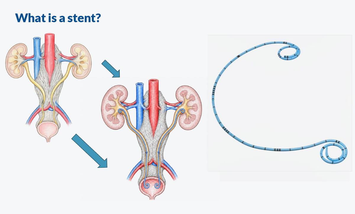
When IgG4-related RPF causes hydronephrosis, teamwork among specialists is crucial. The mechanical restoration of urine flow generally requires the efforts of a urologist (for a ureteral stent) or a radiologist (for a nephrostomy tube)(see below). Simultaneously with these efforts, the rheumatologist, nephrologist, or internist directs the use of immunosuppression, designed to dampen the immune system hyperactivity that has triggered the hydronephrosis.
- Dr. Stuart Wiber, Rheumatologist, Clinical Lecturer, University of Calgary, Department of Medicine
When a stent or nephrostomy tube is needed
A ureteral stent is a small tube placed inside the ureter to help urine move from the kidney to the bladder. It does not treat the immune disease itself, but it can protect the kidney while immune treatment begins to work.
Sometimes the blockage is too tight for a stent to pass. In that situation, a nephrostomy tube may be used. This is a tube placed through the back directly into the kidney to drain urine into a bag outside the body. A nephrostomy tube can feel overwhelming, but it may be kidney-saving when urine cannot drain any other way.
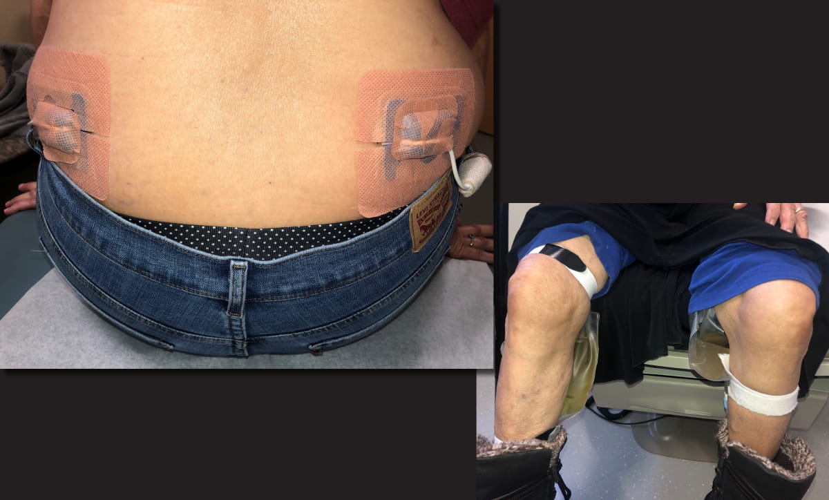
These procedures can be difficult for patients, and the goal is to use stents only for as long as necessary. The ways that patients tolerate stents varies: although some patients have relatively few symptoms, the stents bring daily reminders and considerable discomfort to others. Stents need to be changed over time – generally not less than every six months. Stents often cause pain with certain movements and blood in the urine. They can also predispose to infections.
Sometimes the blockage within a ureter is so tight that the urologist is unable to pass a stent and relieve the obstruction. In such cases, a nephrostomy tube is needed to drain the kidney from another route.
For patients and families, the key message is this: a stent or nephrostomy tube should be a temporary bridge to a more permanent solution. These procedures are designed to protect the kidney while the medical team works on controlling the underlying inflammation.
Dr. Wiber explains how active RPF is treated, including steroids, steroid-sparing medicines, and B-cell therapies that help reduce inflammation and protect organs over time.
Treating active inflammation
Once the flow of urine is restored, the next question is whether the RPF is active and inflamed, mostly scarred and quiet, or a mixture of both.
Glucocorticoids, often called steroids, include medicines such as prednisone that are used to douse inflammation quickly. In IgG4-RD, steroids can reduce swelling and inflammation, especially when treatment starts early. In some cases of IgG4-related RPF, glucocorticoids can lead to clinical and radiographic remission, meaning that the patient’s symptoms, organ function, and imaging studies improve and all signs of active disease are suppressed.1
But steroids are not a perfect answer. They can raise blood sugar, increase blood pressure, weaken bones, cause weight gain, affect mood and sleep, make tissues more fragile, and heighten the risk of infection. Many patients have described steroid-related problems including very high glucose, weight gain, elevated levels of hemoglobin A1c (an indication of poor blood sugar control), blood pressure changes, and concern about bone health.2
For all of these reasons, clinicians try to use steroids carefully. The goal is to quiet inflammation without leaving a person on high doses for too long.
Medicines that can help reduce steroid exposure
Clinicians may also consider “steroid-sparing” medicines. These are treatments that clinicians hope will keep inflammation quiet while reducing the amount of prednisone a person needs over time. Methotrexate is one example of a disease-modifying anti-rheumatic drug, or DMARD.
DMARDs are medicines that calm immune activity more slowly than prednisone but may help maintain disease control as prednisone is tapered. In RPF care, the use of medications such as methotrexate, azathioprine, and mycophenolate may be discussed, depending on the person’s kidney function, other health conditions, and medication risks.
Personalized support on your steroid journey
Get support and education on your steroid journey with a new digital companion, Sam (Steroids and Me). Find answers and learn how to work with your doctors for the best care and taper plan.
Get the appHow B-cell therapy fits in
B cells are immune cells that make antibodies and help activate other immune cells. In IgG4-RD, B cells and their related cells are part of the immune system’s safety team that has become overactive. B-cell therapy aims to quiet that part of the immune response.
Historically, many IgG4-RD specialists have used rituximab, a B-cell-depleting treatment that targets CD20-positive B cells, especially when disease is severe, returns after steroids, or steroids are not safe enough to continue. In RPF, B-cell depletion may be considered when inflammation remains active, when disease returns, or when a person remains dependent on procedures such as stents despite other treatment.
Since 2024, inebilizumab has also become important in IgG4-RD care. Inebilizumab is a CD19-directed B-cell-depleting antibody. Inebilizumab, or Uplizna, is approved by regulatory agencies all over the world for the treatment of IgG4-related disease in adult patients.3
If inflammation is active or likely to return, clinicians may use treatment that calms B-cell activity so the disease is less likely to keep damaging organs.
When monitoring is enough
Not every RPF finding needs immediate immune treatment. Sometimes the tissue looks stable, the kidneys drain well, kidney function is steady, symptoms are mild or absent, and there is no strong sign of active inflammation. In that situation, clinicians may choose watchful monitoring, which means careful follow-up rather than immediate treatment.
Choosing the path of watchful monitoring is not ignoring the disease. Rather, it is an active plan of surveillance that usually includes regular symptom review, blood pressure checks, repeat blood tests, and imaging such as CT, MRI, ultrasound, or PET/CT when appropriate. The medical team watches for signs that the region of RPF is expanding, becoming more inflamed, narrowing a ureter, or affecting kidney function.
This decision is delicate. RPF can be quiet for a time and still need attention later. A patient may feel well even when a ureter is blocked. That is why clinicians do not rely on symptoms alone and intermittent imaging is important.
Surgical intervention considerations
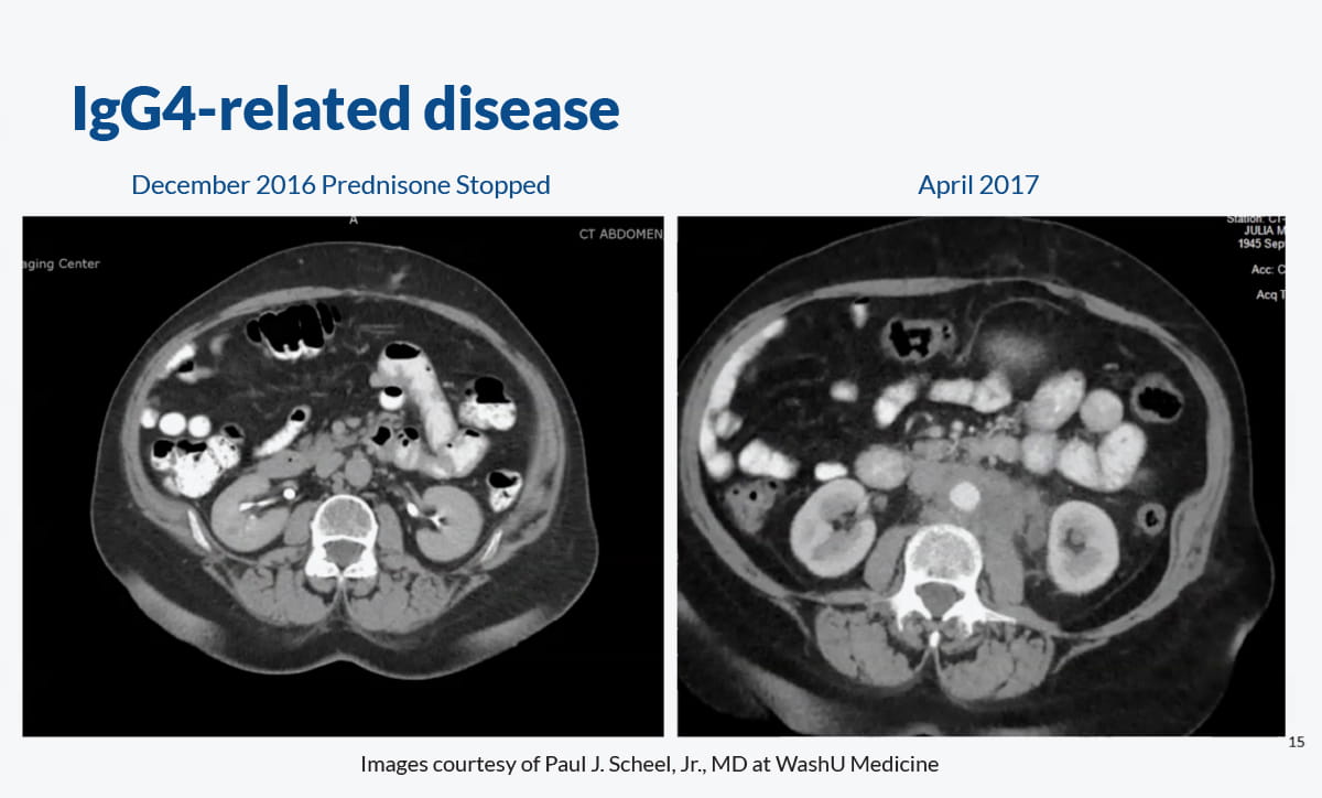
Sometimes in IgG4-related RPF the fibrosis has become so dense that medications, stents, and tubes are all ineffective or undesirable interventions. For example, although medicines may shrink the inflamed tissue to some extent, the ureter(s) remain entrapped by dense fibrosis. In such cases, the problem becomes less like a fire and more like a hardened band around the ureter.
One surgical option is ureterolysis. A ureterolysis procedure, performed under general anesthesia by a urologist, is an attempt to free the ureter from the fibrotic tissue around it. In some cases, the surgeon then moves or secures the ureter in a safer position so it is less likely to be trapped again. A ureterolysis is a delicate procedure requiring great care not to injure the fragile ureteral tissue while freeing it from the surrounding fibrosis.
Ureterolysis may be considered as a later step when inflammation has improved but the ureter remains trapped and a stent still cannot be removed safely. Surgery is not usually the first step when inflammation is active, because medicines may reduce inflammation and make procedures easier or unnecessary. But when a ureter remains mechanically trapped, urology may become central to the care plan again.
What clinicians monitor over time
RPF follow-up is like watching both the road and the engine. Imaging shows the road: whether the tissue is larger, smaller, more active, or stable. Blood and urine tests show the engine: whether the kidneys are keeping up.
Clinicians commonly monitor kidney function with blood tests such as serum creatinine level, cystatic C levels, and the estimated glomerular filtration rate, often called eGFR. The eGFR is a measure that is derived from the patient’s creatinine or cystatin C values as well as their age and sex. They may also check urine tests, non-specific markers of inflammation such as the erythrocyte sedimentation rate (ESR) and C-reactive protein (CRP), serum IgG4 values, and other immune markers.
Blood pressure matters too, because kidney stress can show up as rising blood pressure. A rise in blood pressure can sometimes be a clue that the kidneys are under stress. Follow-up kidney testing and imaging can help the care team see whether RPF is affecting urine drainage or kidney function.
Imaging often plays a central role. Ultrasound can show hydronephrosis. CT or MRI can show the size and location of the retroperitoneal tissue. PET/CT can indicate the degree of inflammation present by measuring metabolic activity within the area.
Active RPF care pathway
Step 1 | Recognize possible RPF |
|---|---|
Step 2 | Ensure adequate kidney drainage and normal flow of urine |
Step 3 | Decide whether inflammation is active |
Step 4 | Treat active disease |
Step 5 | Reassess response |
Step 6 | Address fixed obstruction |
Step 7 | Long-term monitoring |
Questions to bring to an appointment
These questions can help you and your care team make decisions together:
Is my RPF actively inflamed, mostly scarred, or both?
Are one or both ureters narrowed or blocked?
Is there hydronephrosis on imaging?
What are my creatinine and eGFR, and have they changed?
Do I need a ureteral stent or nephrostomy tube to protect kidney function?
What treatment are we using to calm the immune system, and why?
What steroid side effects should we watch for in my case?
Would B-cell therapy be appropriate for me?
How will we know if treatment is working?
When will we repeat imaging?
What would make us consider ureterolysis or another procedure?
What symptoms should make me call urgently?
Summary
Medical management of IgG4-related RPF is really a care pathway, not a single treatment. The first job is to protect the kidneys by making sure urine can drain. The next job is to calm active inflammation, often with steroids, B-cell therapy, or both depending on the situation. If the ureter remains trapped after inflammation improves, urologic procedures may be needed. If the disease is stable and kidney drainage is safe, careful monitoring may be the right choice.
The most reassuring part is that RPF care has become more understood medically. Clinicians now know to look for ureter blockage, kidney function changes, active inflammation, medication risks, and the difference between swelling that may respond to medicine and scar-like tissue that may need a procedure.
Glossary
Retroperitoneal fibrosis (RPF)
Medical definition: Fibro-inflammatory tissue that forms in the retroperitoneum and can surround structures such as the ureters, aorta, and iliac vessels. What it means: Scar-like, inflamed tissue deep in the belly that can wrap around important tubes and blood vessels.
Hydronephrosis
Medical definition: Swelling of the kidney caused by backed-up urine when drainage is blocked. What it means: A kidney “traffic jam” caused by urine that cannot flow out normally.
Ureter
Medical definition: The tube that carries urine from the kidney to the bladder. What it means: The kidney’s drain pipe.
Ureteral stent
Medical definition: A small tube placed inside the ureter to keep urine flowing. What it means: A temporary inner support that helps hold the kidney’s drain pipe open.
Nephrostomy tube
Medical definition: A tube placed through the back into the kidney to drain urine externally. What it means: A backup drain for the kidney when the usual path is blocked.
Glucoroticoids
Medical definition: Anti-inflammatory medicines, such as prednisone, that suppress immune activity. What it means: Strong medicines that turn down the immune system’s fire alarm.
B-cell depletion
Medical definition: Treatment that lowers certain B cells to reduce immune-driven inflammation. What it means: Medicine that quiets part of the immune system’s safety team.
Rituximab
Medical definition: A CD20-directed B-cell-depleting monoclonal antibody used in several immune-mediated diseases and often used off-label in IgG4-RD. What it means: A medicine that lowers certain B cells so inflammation can settle.
Inebilizumab
Medical definition: A CD19-directed B-cell-depleting monoclonal antibody approved for adult IgG4-RD. What it means: A newer B-cell treatment that targets a broader group of B cells.
Ureterolysis
Medical definition: A surgical procedure to free the ureter from surrounding fibrotic tissue. What it means: Surgery to release a trapped kidney drain pipe.
References
Razok A, Romero Noboa ME, Sami F, Patolia KN, Tanveer S. IgG4-related disease and isolated retroperitoneal fibrosis: a narrative review. ARP Rheumatology. 2023;2(2):155-157. https://pubmed.ncbi.nlm.nih.gov/37421192/
IgG4ward Foundation. Understanding Retroperitoneal Fibrosis (RPF) and IgG4-related Disease: Reading Guide. 2025. https://igg4ward.org/media/n2fgjpxg/understanding-igg4rd_rpf_reading-guide.pdf
U.S. Food and Drug Administration. UPLIZNA (inebilizumab-cdon) prescribing information. 2025. https://www.accessdata.fda.gov/drugsatfda_docs/label/2025/761142s004lbl.pdf
Stone JH, Wiber A, and IgG4ward Foundation. Chapter 9 retroperitoneal fibrosis expert video: medical management, kidney protection, stents, immune therapy, procedures, and monitoring. CanJam/IgG4ward educational video.
 Save this lesson so you can resume it later
 Copy Link
 Share On X
 Share On Facebook
 Share On LinkedIn
 Share in Whatsapp
Further reading
IgG4ward reading guide focused on RPF and IgG4-RD. NIH GARD overview of IgG4-related retroperitoneal fibrosis. American College of Rheumatology patient page.Patient guides for doctor appointments
Educating new doctors and healthcare professionals on your IgG4-RD journey can feel like a full-time job. Download, print, or have your doctor scan the QR code of our patient guides to make every appointment more productive.
View guides