
Bookmark this lesson
Login or create an account to save and access your bookmarks
Bookmark this lesson
Login or create an account to save and access your bookmarks
Not yet registered?
What you can access by simply creating a account:
Register today to get the most out of the education academy. Save your progress, bookmarks lessons, track your progress plus more...

Save your progress in this lesson so you can pick it up where you left off.

Add lessons to your bookmarks so you can quickly return to them in the future.

Track your progress throughout the course to see what you have achieved.
Organ affected: Retroperitoneal
IgG4-RD can cause both active inflammation and scar-like fibrosis deep within the abdomen, affecting a variety of important organs.
 6 lessons
 Total: TBC
Organ overview
Understanding the retroperitoneum and how retroperitoneal fibrosis is diagnosed
Retroperitoneal fibrosis, often shortened to RPF, can be a confusing diagnosis because it happens in a part of the body most people have never heard of before. The word retroperitoneal means “behind the peritoneum.” The peritoneum is the thin lining that encloses many organs in the abdomen. So the retroperitoneum is the space that lies behind the peritoneum in the deep back part of the abdomen.
The retroperitoneal space matters because several important structures reside within or pass through it.
The kidneys lie within the retroperitoneal space.
The ureters, which are the small tubes that carry urine from the kidneys to the bladder, run through this area.
Large blood vessels also pass through, including the aorta, which carries blood from the heart to the body.
The inferior vena cava, the largest vein in the body, which carries blood from the lower body back toward the heart, is also here.
When IgG4-related disease affects this area, inflammation can lead to fibrosis, or scar-like tissue. That tissue may form around the aorta, around the ureters, or as a plaque-like area of thickening in the retroperitoneal space.
What symptoms can RPF cause?
RPF can be silent at first. Some people have no symptoms, and the problem is found on a CT scan done for another reason. Others may notice a dull, vague, non-specific pain in the back, side, abdomen, flank, or groin.
Pain may be steady, may wake a person at night, and may be hard to explain. Still other patients present with the abrupt onset of rather severe pain – usually in one of their flanks – that mimics a kidney stone.
Because the ureters pass through the retroperitoneum, RPF can also affect urination. Indeed, if both ureters are affected, a patient may make very little urine because of the impaired flow of urine from the kidney to the bladder.
Blood vessels can also be affected. If the vena cava or other veins are compressed, blood may have trouble returning from the legs to the heart. This can lead to leg swelling or blood clots. If an artery is compressed, a person may notice leg pain while walking that improves with rest. In general, it is more likely that veins rather than arteries will become narrowed, because arteries such as the aorta have thick, muscular walls. The walls of veins are less robust and more subject to compression.
Some people also have fatigue, weight loss, or other symptoms suggesting that the immune system is active and inflammation is present in the body. Such symptoms can be alarming, because they appear to defy simple explanation and can overlap with cancer, infection, kidney disease, spine problems, or other inflammatory conditions.
Area affected | What may happen | Common symptoms or clues |
|---|---|---|
Ureter | Urine flow from the kidney to the bladder may be squeezed or blocked. | Flank or side pain, changes in urination, kidney swelling on imaging, reduced kidney function |
Kidney | Urine backup can put pressure on the kidney. | Fatigue, abnormal kidney blood tests, sometimes few or no symptoms at first |
Aorta or nearby arteries | Blood flow to the legs may be affected. | Leg pain with walking that improves with rest; coldness or aching in the legs |
Vena cava or nearby arteries | Blood return from the legs may be slowed. | Leg swelling, heaviness, sometimes blood clots |
Nearby nerves or soft tissues | Inflammation or fibrosis may irritate nearby tissues. | Back, abdominal, groin, hip, or flank pain |
Systemic (whole-body) inflammation | IgG4-RD activity may affect how a person feels overall. | Fatigue, weight loss, feeling unwell, sometimes abnormal inflammation blood tests |
Watch a video to learn more
Dr. Stuart Wiber, rheumatologist and clinical lecturer at the University of Calgary, explains that when inflammation presses on nearby structures, it can cause real problems: pressure on a ureter can block urine flow and harm the kidney, while pressure on veins or arteries can affect blood flow in the legs.
In this clip, Dr. Stuart Wiber explains the retroperitoneal space and why structures like the ureters, kidneys, aorta, and vena cava are important in IgG4-related retroperitoneal fibrosis.
How doctors narrow down the diagnosis
Diagnosis usually starts with the history. Your care team will ask when symptoms began, where pain is located, whether urination has changed, whether there is leg swelling, and whether you have had IgG4-RD in other organs.
They will also review your previous medical history carefully. This matters because not all RPF is caused by IgG4-RD. Other causes can include non-IgG4 idiopathic RPF, medication reactions, cancer, infection, prior radiation, bleeding, and prior surgery.
“Idiopathic” means doctors have not found a clear cause.
A common patient experience is that the time between the onset of the disease and the establishment of an RPF diagnosis is long. Even if an imaging study identifies thickening or a mass-like swelling in the retroperitoneum, healthcare providers attempting to pinpoint the diagnosis confront significant impediments to easy diagnosis.
How imaging helps
Imaging means pictures of the inside of the body. In RPF, imaging is one of the most important tools.
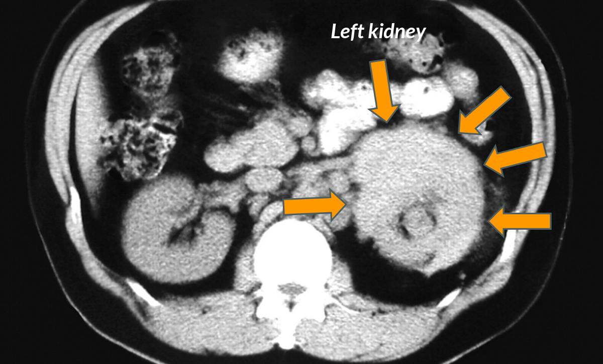
A CT scan can show where the thickened tissue is, whether it surrounds the aorta or ureters, and whether urine is backing up into the kidney.
An MRI may also be used, especially when doctors want a different view of soft tissues or blood vessels.
A PET scan can sometimes help show whether an area appears active with inflammation.
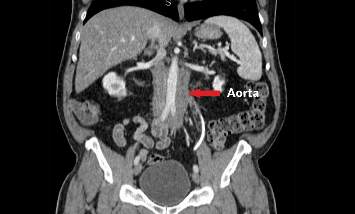
These scans help doctors answer practical questions:
Is the tissue near the aorta or iliac arteries?
Is one or both ureters being squeezed?
Are the kidneys swollen from backed-up urine?
Are blood vessels narrowed or compressed?
Are there signs that another disease may be present?
The words on an imaging report can sound scary. Terms like “mass,” “soft tissue thickening,” “periaortic,” or “fibrosis” do not automatically mean cancer. They mean your care team needs to study the pattern carefully.
What IgG4-RD can look like on imaging
How blood tests help
Blood and urine tests do not diagnose RPF by themselves, but they give important clues. Blood tests permit the monitoring of kidney function and inflammation levels in the body. The following blood tests are frequently useful in RPF and are commonly examined:
Creatinine – a measurement that reflects kidney function. A rising level of blood creatinine reflects worsening kidney function.
C-reactive protein – a general inflammatory marker that is often elevated in RPF.
IgG subclasses – IgG4 is sometimes a useful biomarker.
A high IgG4 level can support the possibility of IgG4-RD, but a normal IgG4 level does not exclude it. In fact, it is notoriously the case that patients with IgG4-related RPF often have misleadingly low or even normal blood concentrations of IgG4. That is why doctors look at IgG4 results together with the full picture, including symptoms, imaging findings, other organ involvement, and sometimes biopsy results.
— Dr. Paul Scheel, Jr., Professor of Medicine and Vice Chancellor for Clinical Affairs at Washington University School of Medicine
When biopsy may be needed
A biopsy is a small tissue sample taken so a pathologist can look at it under a microscope. In IgG4-RD, biopsy can show patterns of inflammation such as the accumulation of immune cells and the development of fibrosis. This can help confirm the diagnosis and rule out mimics such as cancer or infection.
But biopsy in the retroperitoneum is not always simple. The tissue may be close to the aorta, vena cava, ureters, or kidneys – so close that a biopsy may be judged to be not worth the risk of potential complications. Your care team will weigh the benefit of getting tissue against the risk of the procedure.
Sometimes imaging, blood tests, symptoms, organ involvement, and treatment response together give enough information. Other times, biopsy is the safest way to be sure.
When urgent kidney protection is needed
If RPF blocks a ureter, urine may back up toward the kidney. This can cause kidney swelling and kidney injury. In that situation, doctors may bring in a urologist, a specialist who treats the urinary tract.
A urologist may place a stent, which is a small tube that helps keep the ureter open. In some cases, a drainage tube may be placed through the skin into the kidney. These procedures do not treat the immune disease itself, but they can protect the kidney while the care team works on the larger diagnosis and treatment plan.
Why diagnosis takes a team
RPF often needs more than one specialist.
A rheumatologist may guide the IgG4-RD evaluation and immune treatment.
A radiologist reviews imaging patterns.
A urologist helps protect the kidneys and urine flow.
A nephrologist may help if kidney function is affected.
Sometimes vascular, surgical, oncology, infectious disease, or pathology specialists are involved to rule out other causes.
This team approach can feel like a lot of specialists, but it is important for effective care. Because RPF sits near the kidneys, ureters, major blood vessels, and nerves, a team approach helps make sure the right diagnosis is made and the right treatment plan is chosen.
Dr. Scheel explains why diagnosis often depends on teamwork:
“Not only does the pathologist have to be able to read the specimen, there has to be communication between the doctors as to what they’re looking for.”
Summary
The retroperitoneum is the deep back part of the abdomen, where the kidneys, ureters, aorta, vena cava, and nearby nerves are located. In IgG4-RD, inflammation can lead to scar-like tissue in this space. That tissue may press on the ureters, kidneys, or blood vessels and cause symptoms such as back or flank pain, urinary changes, leg swelling, fatigue, or weight loss.
Diagnosis usually combines the full clinical story, imaging, blood and urine tests, and sometimes biopsy. The main goal is to understand whether RPF is related to IgG4-RD, whether another condition could be causing it, and whether any organ—especially the kidney—needs urgent protection.
 Save this lesson so you can resume it later
 Copy Link
 Share On X
 Share On Facebook
 Share On LinkedIn
 Share in Whatsapp
Further reading
IgG4ward! reading guide on retroperitoneal fibrosis and IgG4-RD. NIH GARD overview of IgG4-related retroperitoneal fibrosis. IgG4ward! Academy Chapter 1: What is IgG4-RD?Patient guides for doctor appointments
Educating new doctors and healthcare professionals on your IgG4-RD journey can feel like a full-time job. Download, print, or have your doctor scan the QR code of our patient guides to make every appointment more productive.
View guides