
IgG4ward! EuroJAM Session 6: Retroperitoneal fibrosis, back pain, stents, and kidney health
Learn how retroperitoneal fibrosis can affect people living with IgG4-related disease. Dr. Archie Hughes-Hallett and Joanna Teles, a Kidney Nurse Specialist, explain back and abdominal pain, urinary obstruction, hydronephrosis, ureteric stents, nephrostomy tubes, surgical considerations, and kidney health.

Retroperitoneal fibrosis can surround the ureters, obstruct the flow of urine, and place kidney function at risk. Treatment may include controlling the underlying inflammation and relieving the obstruction with a ureteric stent, nephrostomy tube, or, in selected cases, surgery.
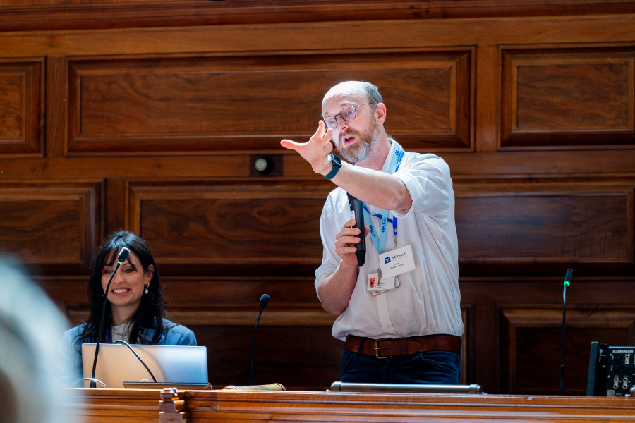
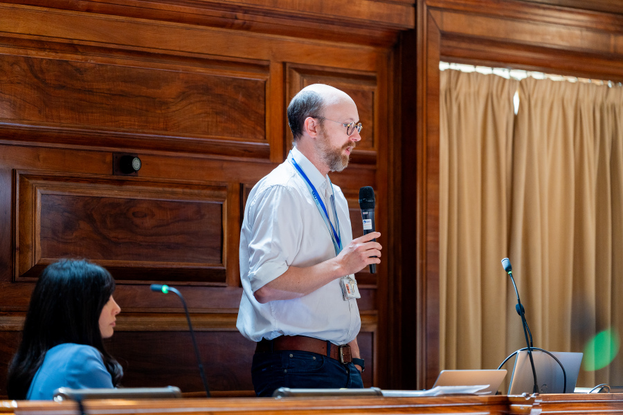
In Session 6 of the IgG4ward! EuroJAM Educational Series, Dr. Archie Hughes-Hallett, Consultant Urologist at Imperial College Healthcare NHS Trust, and Joanna Teles, Kidney Nurse Specialist at Imperial College Healthcare NHS Trust, explain retroperitoneal fibrosis using accessible language, anatomical illustrations, and a patient case example.
Retroperitoneal fibrosis, or RPF, occurs when inflammation and scar tissue develop in the area behind the lining of the abdomen. This tissue can surround structures in the abdomen and place pressure on the ureters, the tubes that carry urine from the kidneys to the bladder.
When urine cannot flow normally through a ureter, it may build up in the kidney. This is called hydronephrosis. If the obstruction is not relieved, it can affect kidney function.
The speakers discuss how ureteric stents and nephrostomy tubes may be used to relieve obstruction and protect the kidneys. They also explore longer-term approaches, including regular stent changes and ureterolysis, an operation that frees the ureters from surrounding scar tissue.
Audience questions and shared experiences are woven throughout the session, creating a patient-centered conversation about pain, diagnosis, imaging, medical treatment, surgery, kidney health, and shared decision-making.
Watch the session:
Key Takeaways from IgG4ward! EuroJAM Session 6
Explore the questions below for key information discussed by Dr. Archie Hughes-Hallett, Joanna Teles, and members of the EuroJAM community during this interactive session.
Retroperitoneal fibrosis, or RPF, occurs when inflammation and scar tissue develop in the retroperitoneum, the area behind the membrane lining the abdomen. This tissue commonly forms around the aorta and may surround or compress nearby structures, including the ureters.
Retroperitoneal fibrosis can be a manifestation of IgG4-related disease. However, RPF can also occur without a clearly identified cause, which may be described as idiopathic retroperitoneal fibrosis. The speakers explain that the conditions can overlap significantly and may behave similarly from a urological perspective.
The ureters are narrow tubes that carry urine from the kidneys to the bladder. In retroperitoneal fibrosis, inflammation and scar tissue can surround or compress one or both ureters, preventing urine from draining normally. If the obstruction is not relieved, it can affect kidney function.
Hydronephrosis is swelling of the kidney caused by a buildup of urine. In retroperitoneal fibrosis, it may develop when scar tissue narrows or blocks a ureter and causes urine to flow back toward the kidney.
The session explains that severe obstruction may cause a sudden change in kidney function, while longer-term obstruction can contribute to chronic kidney disease.
Retroperitoneal fibrosis may cause pain in the back, flank, abdomen, or surrounding areas. The pain may be persistent, dull, or difficult to pinpoint.
Because back pain is common and can have many causes, symptoms alone may not reveal whether RPF is responsible. Imaging and clinical evaluation can help the healthcare team investigate the cause.
A ureteric stent or nephrostomy tube may be used to relieve an obstruction and protect kidney function. Medical treatment may also be used to address the underlying inflammation.
The appropriate approach depends on the individual situation, including the severity of the obstruction, kidney function, symptoms, imaging, and response to treatment.
A ureteric stent is a long, flexible tube placed inside the ureter. One end sits in the kidney and the other in the bladder.
The stent helps urine move from the kidney to the bladder despite pressure or narrowing affecting the ureter. Some people need stents temporarily, while others may live with them longer and have them changed periodically.
Some people experience few problems with a stent, while others may have pain, urinary frequency, discomfort, or blood in the urine.
Dr. Hughes-Hallett explains that a sudden worsening of symptoms, fever, burning when passing urine, or feeling generally unwell may indicate a problem such as infection or a blocked stent. These changes should be discussed promptly with a healthcare professional.
A nephrostomy is a tube inserted through the back directly into the kidney. It allows urine to drain without passing through the ureter.
A nephrostomy may be needed when the ureter is severely obstructed, when a stent cannot be placed, or when a stent is no longer relieving the obstruction adequately.
Ureterolysis is an operation that carefully frees the ureters from surrounding scar tissue and moves them away from the affected area.
The procedure may help some people become free from stents or nephrostomy tubes. However, it is a significant operation with potential risks and is not appropriate for everyone. The session explains why the possible benefits and risks must be considered for each individual.
Some anti-inflammatory pain medications, including certain medications available without a prescription, can affect kidney function when used regularly.
Joanna encourages patients to tell their healthcare team about ongoing pain and any pain medications they are using. This allows the team to consider the person’s kidney function and discuss appropriate alternatives when needed.
The session highlights hydration, blood pressure management, a balanced diet, physical activity, avoiding smoking, and maintaining a healthy weight as important parts of supporting kidney and overall health.
The speakers also encourage patients to write down their most important questions before an appointment. Understanding the available options, including their possible benefits and risks, can help patients participate in decisions about their care. If something is unclear or a question has not been answered, patients are encouraged to ask again.
Moments from the EuroJAM Session 6:





IgG4ward! EuroJAM Session Series
This presentation is the sixth release in the IgG4ward! EuroJAM Educational Series, which brings expert presentations and patient-centered conversations from EuroJAM to the broader IgG4-RD community.
Subscribe to the IgG4ward! Foundation YouTube channel and follow the Foundation to explore additional sessions and future educational programs.
Medical Disclaimer - Educational Purposes Only
The information provided on this page and in the accompanying EuroJAM session is intended for educational and informational purposes only and is not intended to serve as medical advice, diagnosis, or treatment.
This content reflects key themes discussed during Dr. Archie Hughes-Hallet and Joanna Teles's EuroJAM session.
Individuals should always consult their physician or qualified healthcare provider regarding questions about their medical condition, symptoms, kidney health, medications, diagnosis, treatment options, or healthcare decisions.
Never disregard professional medical advice or delay seeking medical care because of information provided on this page or in the accompanying educational resources.
Thank you to our Industry Sponsors
The IgG4ward! Foundation gratefully acknowledges the organizations whose support helped make IgG4ward! EuroJAM possible.
All educational content presented during the event was developed independently by the IgG4ward! Foundation and its invited speakers.




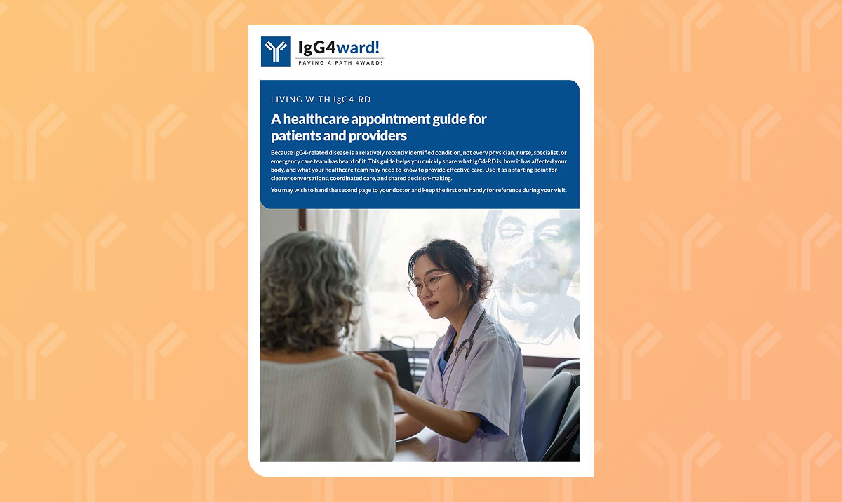
A healthcare appointment guide for patients and providers
A guide to help you quickly share what IgG4-RD is, how it has affected your body, and what your healthcare team may need to know to provide effective care.
Read more
What to expect at IgG4ward! CamJAM: October 11 in Cambridge
Learn what to expect at IgG4ward! CamJAM on October 11, 2026, in Cambridge, Massachusetts: IgG4-RD education, expert Q&A, and connection for patients and caregivers.
Read more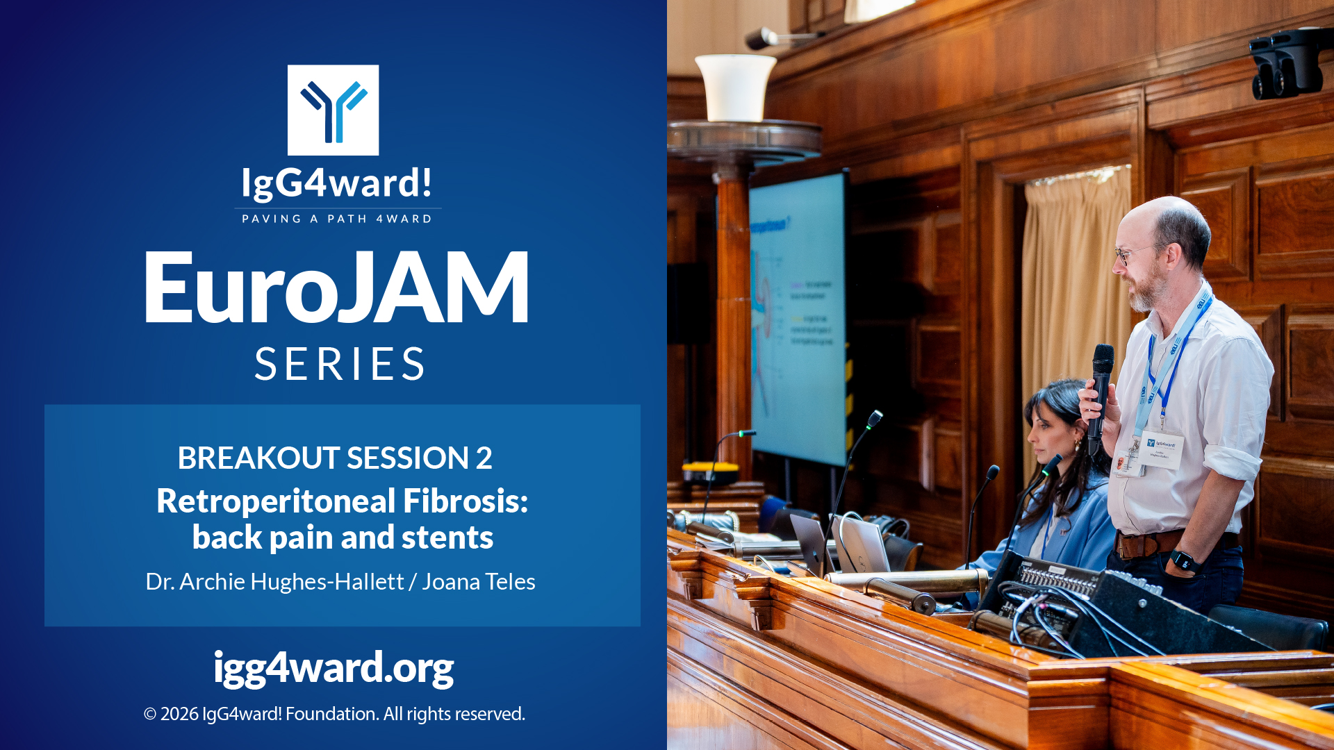
IgG4ward! EuroJAM Session 6: Retroperitoneal fibrosis, back pain, stents, and kidney health
Learn how retroperitoneal fibrosis can affect people living with IgG4-related disease. Dr. Archie Hughes-Hallett and Joanna Teles, a Kidney Nurse Specialist, explain back and abdominal pain, urinary obstruction, hydronephrosis, ureteric stents, nephrostomy tubes, surgical considerations, and kidney health.
Read more
IgG4ward! EuroJAM: Hope in action
Hope was a theme throughout the IgG4ward! EuroJAM - but not as wishful thinking. Patients, caregivers, researchers, physicians and industry partners came together to share knowledge, build connections and move the conversation around IgG4-related disease forward. Here's a look back at the event and what's coming next.
Read more